1. Key super-resolution platforms
MAxSIM — multi-angle-crossing structured illumination with height-controlled mirror (custom) Real-time, live-cell super-resolution to map plasma-membrane topology and 3D single molecule tracking (3D-SMT) in near real-time (0.5-1 Hz) in live cells. Quantifies curvature and domain enrichment during stress adaptation and invasive transitions.
SIM — Structured Illumination Microscope (custom) Fully customizable 2D/3D, 4-color super-resolution workflows. Used to resolve membrane domains and receptor co-localization at the nanoscale.
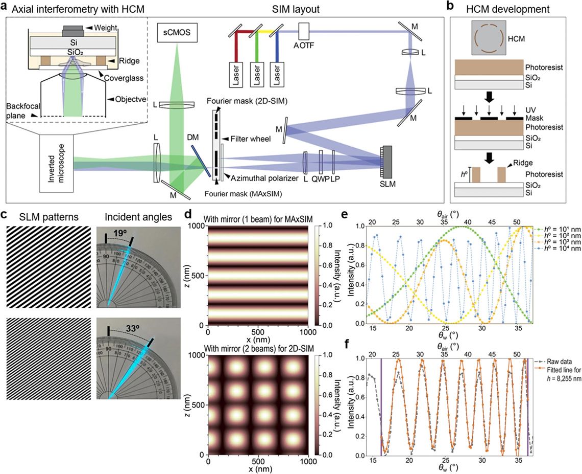
MAxSIM layout and axial interferometry
2. Live-cell imaging & photomanipulation
Spinning-Disk Confocal High-speed volumetric imaging with environmental control for live cells. Ideal for tracking cytoskeletal remodeling and rapid signaling responses.
Total Internal Reflection Fluorescence (TIRF) and single molecule tracking Selective imaging of the ~100–200-nm membrane–glass interface for single-molecule recruitment, exo/endocytic events, and receptor trafficking at the plasma membrane.
FRAPPA photomanipulation module ROI-targeted photobleaching/photoactivation for diffusion, turnover, and trafficking kinetics; integrates with confocal and TIRF modalities.
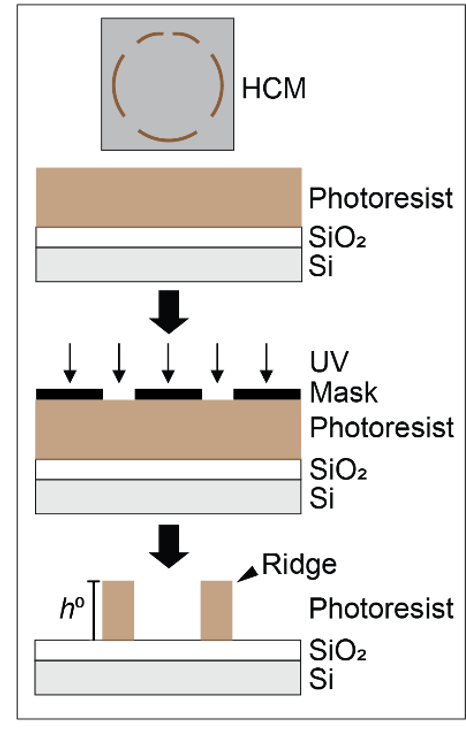
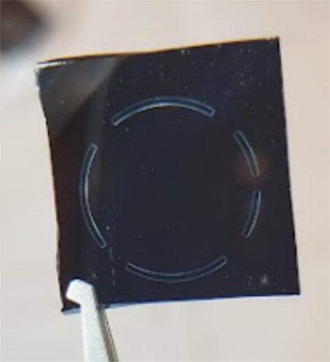
Height-controlled mirror (HCM) fabrication
3. Mechanics & biophysics measurements
Nanoindenter (Optics11 Chiaro) Sub-nanoNewton force measurements of cells and tissues to quantify elastic modulus, cortical stiffness, and viscoelastic responses during mechanical stress.
4. AI & analysis pipelines
AI & Computational Pipelines CNN-based pathology classification of IHC and H&E image trained DCIS risk stratification and custom MAxSIM analysis.
MAxSIM-enabled 3D single molecule tracking (SMT)
2D-structured illumination microscopy